The Electrocardiogram (ECG or EKG) is a recording of the heart’s electrical activity from the surface of the body. Electrical activity is generated in the heart by the movement of ions across cardiac cell membranes during muscle contraction.
The first ECG on a dog was performed in 1887 by Augustus Waller on his pet Bulldog ‘Jimmy’. Thankfully, today we do not require our patients to stand in pots of seawater to obtain an accurate ECG!
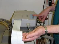
An ECG is generally performed without sedation or a general anaesthetic. ECG clips are placed on the skin over the elbow and stifle joints. The clips are then sprayed with surgical spirit to improve electrical conduction. An ECG is then obtained over a 2-3 minute period.


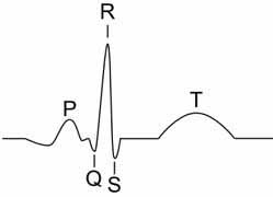
P wave This represents atrial contraction
PR interval Represents a delay in conduct of the electrical impulse between the atria and ventricles caused by the atrioventricular node
QRS complex Represents the contraction of the ventricles
T wave Represents relaxation of the ventricles

Why might we suggest an ECG
Indications of obtaining an ECG include:
- History of collapse or fainting
- Detection of a heart arrhythmia during examination
- Investigation into heart disease
- Monitoring during anaesthesia or in a critical care patient


